


About Service
Interventional neuroradiology is a medical unit that diagnoses and treats vascular diseases of the brain, spinal cord and nervous system using imaging techniques. These procedures, performed using minimally invasive methods, are carried out inside the blood vessels using imaging techniques such as angiography, MRI and CT.
Conditions such as brain aneurysms, arteriovenous malformations (AVMs), stroke, and vascular occlusions are treated without the need for surgical incisions. Methods such as embolisation, recanalisation, and diagnostic procedures shorten recovery times and reduce risks.
What is Interventional Neuroradiology?
Interventional neuroradiology is a minimally invasive method in which vascular diseases of the brain, spinal cord and nervous system are treated with imaging techniques. Vessels are examined using methods such as angiography, MRI and CT, and treatment is performed directly inside the vessel.
A thin catheter inserted through the groin is used to reach the problematic vessel and administer treatment. This allows conditions such as brain aneurysms, vascular occlusions, arteriovenous malformations (AVMs), and strokes to be treated without the need for surgical incisions. These procedures, which are less traumatic than surgical methods, enable patients to recover more quickly.
What Conditions Does Interventional Neuroradiology Treat?
Interventional neuroradiology plays an important role in the diagnosis and treatment of vascular diseases of the brain, spinal cord, and nervous system. This minimally invasive technique, performed inside the blood vessel with the help of imaging techniques, allows many neurovascular diseases to be treated without the need for surgery. It is an effective treatment option, especially in serious conditions such as sudden vascular occlusions, aneurysms, and vascular disorders.
The diseases treated by interventional neuroradiology are as follows:
| Disease | Explanation |
| Brain Aneurysm | It is performed to close balloon-shaped dilations that form in the blood vessels of the brain. |
| Arteriovenous Malformation (AVM) | It is used in the treatment of abnormal connections in the blood vessels of the brain. |
| Acute Stroke (Paralysis) | Blood flow is restored by removing the clot from the brain vessel (thrombectomy). |
| Vascular Occlusions | The narrowing or blockages in the blood vessels of the brain and neck are opened up. |
| Dural Arteriovenous Fistula (dAVF) | It closes abnormal connections between the blood vessels in the brain membrane. |
| Carotid and Vertebral Artery Stenosis | It enables the treatment of narrowing in the neck arteries using methods such as stenting. |
| Cerebral haemorrhage | It is used to close vascular defects that cause bleeding. |
| Tumour Embolisation | Reducing blood flow is achieved by blocking the vessels that feed brain tumours. |
| Spinal Vascular Malformations | It is used in the treatment of abnormalities in the spinal cord vessels. |
| Epistaxis (Severe Nose Bleed) | The artery causing the bleeding is closed by means of embolisation. |
Treatment Methods Used in Interventional Neuroradiology
Interventional neuroradiology offers minimally invasive methods for the diagnosis and treatment of brain and spinal cord vascular diseases. These procedures, performed inside the blood vessels with the assistance of imaging techniques, enable the treatment of diseases without the need for surgical intervention.
Treatment methods used in interventional neuroradiology are primarily categorised into three main groups:
- Embolisation (blockage),
- Recanalisation (opening of blood vessels),
- Diagnostic procedures.
Embolisation Procedures
Embolisation procedures are treatment methods that aim to close abnormal or diseased blood vessels. During these procedures, blood flow is stopped by placing special materials inside the vessel, thereby isolating the diseased area. Embolisation is used both to reduce the risk of bleeding and to treat diseased tissues.
The main embolisation procedures performed in interventional neuroradiology are as follows:
- Brain Aneurysm Embolisation: Ruptured or unruptured brain aneurysms are closed using coils, stents or flow-diverting devices.
- Arteriovenous Malformation (AVM) Treatment: Abnormal connections in the brain vessels are blocked using the embolisation method to prevent blood flow.
- Dural Arteriovenous Fistula Treatment: Abnormal connections between the vessels in the brain membrane are treated by embolisation.
- Carotid-Cavernous Fistula Treatment: Abnormal connections between arteries and veins are closed.
- Spinal AVM/AVF Treatment: Abnormal connections in spinal cord blood vessels are treated using the embolisation method.
- Intracranial Tumour Embolisation: The vessels supplying meningioma and other intracranial tumours are blocked to shrink the tumour.
- Epistaxis (Nose Bleed) Embolisation: In severe nose bleeds, the vessels causing the bleeding are closed.
- Traumatic Vascular Injuries: Intravascular treatment methods are used in cases of carotid or vertebral artery injuries.
Recanalisation (Vessel Opening) Procedures
Recanalisation procedures are performed to reopen blocked or narrowed vessels and restore normal blood flow. These methods are particularly vital in cases of stroke and vascular occlusion. Recanalisation enables sufficient blood flow to the brain or other tissues through blocked vessels.
The recanalisation methods used in interventional neuroradiology are as follows:
- Acute Stroke Treatment (Clot Removal - Thrombectomy): This is the mechanical removal of the clot blocking the brain artery.
- Carotid and Vertebral Artery Stenting: Stents are placed to open narrowings in the neck and spinal arteries.
- Intracranial Artery Stenting: Stents are placed in narrowed arteries within the brain to regulate blood flow.
- Subclavian Artery Stenting: Stents are applied to open narrowings in the subclavian artery, which carries blood to the arm and brain.
Diagnostic Procedures
Diagnostic procedures are performed to obtain detailed images of the blood vessels in the brain and spinal cord. These procedures enable the accurate diagnosis of vascular diseases and the planning of treatment. During imaging, special contrast agents are administered into the blood vessels to obtain detailed images.
The main diagnostic procedures used in interventional neuroradiology are as follows:
- Digital Subtraction Angiography (DSA): Access is gained through the groin or wrist artery to provide detailed imaging of the brain and spinal cord vessels.
- Arcus, Thoracic and Abdominal Aortogram: Used to visualise the large vessels emerging from the heart and the surrounding vascular structures.
- Inferior Petrosal Sinus Sampling: A special imaging and sampling method used in the diagnosis of Cushing's syndrome.
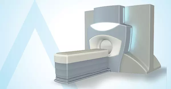
Imaging Methods Used in Interventional Neuroradiology
Accurate and detailed imaging is crucial for the success of interventional neuroradiology procedures. Imaging methods allow for detailed examination of the brain, spinal cord, and vascular structures, enabling the identification of diseased areas. These techniques play an important role in both diagnosis and guiding the treatment process.
Magnetic Resonance Angiography (MRA)
Magnetic Resonance Angiography (MRA) is a magnetic resonance (MR) technique that provides detailed imaging of vascular structures. The internal structure of blood vessels can be clearly visualised using contrast agents. MRA is particularly preferred for detecting aneurysms, vascular occlusions, and arteriovenous malformations in the brain and neck vessels. It is a safe method as it does not involve radiation.
Computed Tomography Angiography (CTA)
Computed Tomography Angiography (CTA) is a tomography method used to obtain cross-sectional images of blood vessels. A contrast agent is administered intravenously to produce a detailed map of the blood vessels. It is frequently used in the evaluation of aneurysms, vascular stenoses, and occlusions. The procedure is rapid and provides the benefit of rapid diagnosis in emergency situations (e.g., suspected stroke).
Digital Subtraction Angiography (DSA)
Digital Subtraction Angiography (DSA) is the gold standard imaging method that provides the clearest images of the brain and spinal cord vessels. Contrast material is administered via the groin or wrist artery, and the vascular structures are visualised in real time. It is widely used in the diagnosis of aneurysms, AVMs, vascular occlusions, and fistulas. Furthermore, many interventional treatment procedures are performed under DSA guidance.
Recovery Process Following Interventional Neuroradiology Procedures
As interventional neuroradiology procedures are performed using minimally invasive techniques, the recovery process is generally rapid and comfortable. These procedures are less traumatic than surgical interventions, allowing patients to return to their daily lives within a short period. The recovery process may vary depending on the treatment administered, the patient's general health, and post-procedure care. However, most patients can resume normal activities within a few days.
The following should be noted during the post-procedure recovery process:
- The patient remains under observation for several hours after the procedure.
- A pressure bandage is applied to the entry site in the groin or wrist area.
- Avoid strenuous physical activity for the first 24 hours.
- The entry site should be checked regularly.
- The use of blood-thinning medication is regulated according to the doctor's recommendation.
- Adequate fluid intake is recommended.
- In case of dizziness or severe headache, a doctor should be consulted.
- The doctor should be informed if redness, swelling or bleeding occurs at the entry site.
- Regular doctor check-ups should not be neglected.
- The doctor's recommendations should be followed throughout the healing process.
Interventional Neuroradiology Risks and Precautions
Although interventional neuroradiology procedures are minimally invasive, they may involve certain risks. While the procedures are generally considered safe, some undesirable situations may arise due to the intravascular pathways used and the techniques applied. To minimise risks, the procedures should be performed by experienced specialists, and the patient should be informed about the pre- and post-procedure processes. Furthermore, patients' careful attention after the procedure positively affects the recovery process.
Possible risks and precautions related to the procedure include:
- Bruising or slight bleeding may occur at the insertion site.
- In rare cases, a blood clot may form in the vessel.
- An allergic reaction to the contrast medium may develop.
- Although the risk of infection is low, the insertion site should be kept clean.
- Mild headache or fatigue may be felt after the procedure.
- Swelling or pain may occur at the insertion site.
- Heavy activities should be avoided for a few days after the procedure.
- Drinking plenty of fluids helps eliminate the contrast material.
- A doctor should be consulted in case of sudden dizziness, loss of vision or severe pain.
- Regular doctor check-ups should not be neglected.
Frequently Asked Questions About Interventional Neuroradiology
How Are Interventional Neuroradiology Procedures Performed?
The procedures are generally performed under local anaesthesia, using thin catheters inserted through a vein in the groin. With the help of imaging techniques, the problematic vessels are targeted and treated.
Are Interventional Neuroradiology Procedures Painful?
Most interventional neuroradiology procedures are performed under local anaesthesia, so the patient does not feel pain during the procedure. In some cases, sedation or general anaesthesia may also be preferred.
What is the Recovery Process After the Procedure?
The recovery process after interventional neuroradiology procedures is generally rapid. Patients can often return to their daily lives within a few days.
How Long Do Interventional Neuroradiology Procedures Take?
The duration of the procedure varies depending on the condition being treated, but it is usually completed within 1 to 3 hours. The duration may be longer in more complex cases.
Who Can Undergo Interventional Neuroradiology Treatment?
Patients with neurovascular diseases who are not suitable for surgical treatment can be treated with this method. The patient's suitability is determined following the doctor's evaluation.
Is Interventional Neuroradiology Safe?
When performed by teams of specialists in the field, interventional neuroradiology procedures are highly safe. Thanks to modern imaging techniques, procedures are performed with precision.
How Should One Prepare Before Interventional Neuroradiology Procedures?
Before the procedure, the patient may need to fast and discontinue certain medications under the doctor's supervision. Blood tests and imaging examinations are also performed.
What is the Difference Between Interventional Neuroradiology and Surgical Treatment?
Interventional neuroradiology is a minimally invasive method, and treatment is administered without making large incisions in the body. This results in a shorter recovery time and reduced hospital stay.